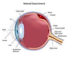
CEN Retinal Detachment
CEN Retinal Detachment
Retinal detachment is a disorder of the eye in which the retina detaches from the retinal pigment epithelium. Retinal detachments can be caused by fluid leaking behind the retina through tears, by traction on the retina, or by fluid exuding from the retina. The visual prognosis of a retinal detachment is dependent on the duration of the detachment, whether the macula was detached, and the underlying health of both the retina and circulatory system of the eye.
The retina is a thin layer of light sensitive tissue on the back wall of the eye. The optical system of the eye focuses light on the retina much like light is focused on the film or sensor in a camera. The retina translates that focused image into neural impulses and sends them to the brain via the optic nerve. Occasionally, posterior vitreous detachment, injury or trauma to the eye or head may cause a small tear in the retina. The tear allows vitreous fluid to seep through it under the retina, and peel it away like a bubble in wallpaper.
Signs and Symptoms
A rhegmatogenous retinal detachment is commonly preceded by a posterior vitreous detachment which gives rise to these symptoms:
- flashes of light (photopsia) – very brief in the extreme peripheral (outside of center) part of vision
- a sudden dramatic increase in the number of floaters
- a ring of floaters or hairs just to the temporal (skull) side of the central vision
Although most posterior vitreous detachments do not progress to retinal detachments, those that do produce the following symptoms:
- a dense shadow that starts in the peripheral vision and slowly progresses towards the central vision
- the impression that a veil or curtain was drawn over the field of vision
- straight lines (scale, edge of the wall, road, etc.) that suddenly appear curved (positive Amsler grid test)
- central visual loss
In the event of an appearance of sudden flashes of light or floaters, an eye doctor needs to be consulted immediately. A shower of floaters or any loss of vision, too, is a medical emergency.
Risk Factors
The following factors increase your risk of retinal detachment:
- Aging — retinal detachment is more common in people over age 50
- Previous retinal detachment in one eye
- A family history of retinal detachment
- Extreme nearsightedness (myopia)
- Previous eye surgery, such as cataract removal
- Previous severe eye injury
- Previous other eye disease or inflammation
Diagnosis
- Retinal examination – The doctor may use an instrument with a bright light and a special lens (ophthalmoscope) to examine the back of your eye, including the retina. The ophthalmoscope provides a highly detailed view, allowing the doctor to see any retinal holes, tears or detachments.
- Ultrasound imaging – this test if bleeding has occurred in the eye, making it difficult to see your retina.
CEN Retinal Detachment – Types
- Rhegmatogenous retinal detachment – A rhegmatogenous retinal detachment occurs due to a break in the retina (called a retinal tear) that allows fluid to pass from the vitreous space into the subretinal space between the sensory retina and the retinal pigment epithelium. Retinal breaks are divided into three types – holes, tears and dialyses. Holes form due to retinal atrophy especially within an area of lattice degeneration. Tears are due to vitreoretinal traction. Dialyses are very peripheral and circumferential, and may be either tractional or atrophic. The atrophic form most often occurs as idiopathic dialysis of the young.
- Exudative, serous, or secondary retinal detachment – An exudative retinal detachment occurs due to inflammation, injury or vascular abnormalities that results in fluid accumulating underneath the retina without the presence of a hole, tear, or break. In evaluation of retinal detachment it is critical to exclude exudative detachment as surgery will make the situation worse, not better. Although rare, exudative detachment can be caused by the growth of a tumor on the layers of tissue beneath the retina, namely the choroid. This cancer is called a choroidal melanoma.
- Tractional retinal detachment – A tractional retinal detachment occurs when fibrous or fibrovascular tissue, caused by an injury, inflammation or neovascularization, pulls the sensory retina from the retinal pigment epithelium.
Treatment
- Cryopexy and laser photocoagulation
- Scleral buckle surgery
- Pneumatic retinopexy
- Vitrectomy
Emergency Room Certification Courses
Overview
- Elite Reviews Offers A Variety Of Online Courses That Will More Than Adequately Help Prepare The Emergency Nurse To Pass The National Exam.
- Each Course Includes Continuing Education Credit and Sample Questions.
Continuing Education
- Each Of Our Online Courses Has Been Approved Continuing Education Contact Hours by the California Board of Nursing
- Login To Your Account In Order To Access The Course Completion Certificate Once The Course Is Complete.
CEN Free Trial
- FREE Sample Lecture & Practice Questions
- Available For 24 Hrs After Registration
- Click Free Trial Link To Get Started – CEN Free Trial
How It Works
How The Course Works
- First – Purchase The Course By Clicking On The Blue Add To Cart Button – You Will Then Be Prompted To Create A User Account.
- Second – After Creating An Account, All 3 Options (90, 120 or 150 Days) Will Be Listed. Select The Option You Desire And Delete The Other Two.
- Third – You Will Be Prompted To Pay For The Review Using PayPal – After Payment You Will Be Redirected Back To Your Account.
- Last – Click The Start Button Located Within Your Account To Begin The Program
- 175 Sample Questions
- Q & A With Rationales
- Approved For 5 CEU’s
- 90 Days Availability
- Cost $75.00
- 1250+ Sample Questions
- Q & A With Rationales
- Approved For 25 CEU’s
- 90 Days Availability
- Cost $200.00
CEN Practice Questions Bundle
- 1350+ Sample Questions
- Q & A With Rationales
- Approved For 30 CEU’s
- 90 Days Availability
- Cost $225.00
CEN Review Course
- Option 1
- Lectures & 1250+ Questions
- Approved For 35 CEU’s
- 90 Days Availability
- Cost $325.00
- Option 2
- Lectures & 2000+ Questions
- Approved For 40 CEU’s
- 90 Days Availability
- Cost $350.00
CEN Review Course Bundle
- Option 3
- Lectures & 3000+ Questions
- Approved For 70 CEU’s
- 90 Days Availability
- Cost $375.00


